Illustrated Medicine: Case Study
Trimalleolar Fracture-Dislocation
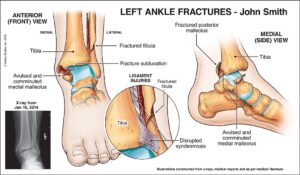
Incident
This 21-year-old construction worker slipped into a hole, sustaining a displaced/dislocated trimalleolar fracture that required open reduction internal fixation (ORIF). Osteoarthritis (OA) developed quickly in the joint due to the impact to, and disruption of, the articular cartilage.
Purpose of illustration
This panel was created to demonstrate the significant fracture-dislocation with associated injury to the syndesmosis (the distal tibia-fibula joint bound together by ligaments). The illustrations allowed litigation participants to fully appreciate the severity of the trauma and why OA ensued.
Rationale for litigation visual
Although x-rays demonstrated the fracture-dislocation, illustrations were required to translate the films into more readily understood images to show the full extent of trauma, including details of the syndesmosis injury.
Complications
Post-injury, this young man suffered pain on ambulation and at rest. His ankle joint demonstrated osteoarthritic degeneration within a year of the slip & fall. Some of the hardware and osteophytes required surgical removal. Further degeneration and associated surgeries were predicted for him.
Other visuals
Additional illustrations showed: the post-op appearance after ORIF surgery; hardware removal; future further osteoarthritic degeneration; and potential future ankle surgery – including debridement, fusion or replacement.
Outcome
This exhibit was created to allow the orthopedic expert to fully explain the extent of trauma this man sustained, and the significance of his intra-articular fractures. The illustrations were sent to defense and the case settled before trial.