Illustrated Medicine: Case Study
Double puncture of colon during gallbladder surgery –
Case settles prior to trial
Incident
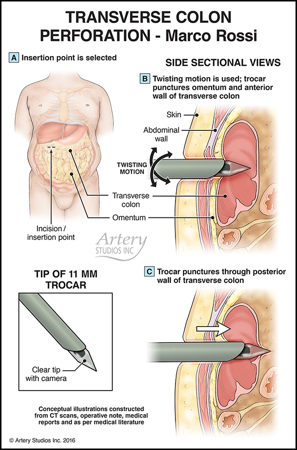
While undergoing cholecystectomy surgery, the anterior and posterior walls of Mr. Rossi’s transverse colon were perforated, leading to fecal peritonitis, adhesion formation and incarcerated (trapped) umbilical hernia.
Purpose of illustration
This exhibit was created to demonstrate the mechanism of injury to Mr. Rossi’s colon – as per the findings upon surgical reopening, associated imaging and the case medical reports.
Rationale for litigation visual
This illustration clarifies the complex medical terminology and difficult-to-understand radiology. It provides a clear view of what occurred during the initial surgery.
Complications
Brown fluid was observed in Mr. Rossi’s abdominal drains, and intra-abdominal air was seen on CT – indicative of a perforated bowel. He required surgical repair of the perforations, removal of pus and omentum from his abdominal hernia, and a colostomy. Later, surgical reversal of his colostomy site was undertaken along with repair of the abdominal hernia. He developed a pulmonary embolism after this surgery, resulting in his death.
Other visuals demonstrated
Additional exhibits demonstrated: the anatomy of the abdomen and intestinal tract; enlargement of Mr. Rossi’s hernia (as per CT imaging); as well the numerous surgeries performed.
Outcome
The illustrations were used at a settlement conference and the case resolved close to the doctor’s policy limits.
“Working with Artery Studios was instructive because of their knowledge of anatomy and how to illustrate it … and skills and capacity to demonstrate tissues, planes, and perspective. The images helped my understanding and to feel more confident in what we could present to a jury … and that positively impacted our settlement negotiations.” – James Coogan, Dwyer & Coogan, PC