Illustrated Medicine: Case Study
Intra-Articular Wrist Fracture
Incident
Mr. Jones was a 45-yo male driver involved in a head-on collision and required extrication.
Purpose of Illustration
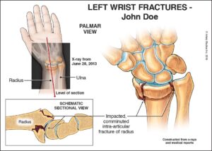
This panel was created to show details of his left wrist injury, providing a visual translation of the radiology and medical reports. It also served to demonstrate the severity of the trauma-related issue(s), to address objections of pre-existing psoriatic arthritis and two prior MVA’s with unrelated sequelae.
Rationale for litigation visual
X-rays were too difficult for a jury to comprehend understand. Other visuals showed the surgery performed and degeneration concepts resulting from the significant damage he sustained to the articular cartilage in his wrist.
Complications
He was later shown to have early arthritic changes in his left wrist joints – based on x-ray findings and a bone scan. Experts opined that his wrist would likely continue to degenerate with osteoarthritis.
Other visuals demonstrated
Concepts of: concussion; lumbar spine injury/surgery; pain treatments; sciatica complications; and right foot issues including fractures, surgeries, and degeneration. A 3D-printed model showed details of his right foot fractures.
Outcome
The demonstrative evidence was created in time for mediation. Using the visuals, the lawyer walked through the medical issues in detail, linking them directly to the biomechanical engineering opinions. A significant settlement was reached shortly thereafter with the visuals showing the clear “facts” and informing the adjuster’s decision.