Shoulder Injury from Head-on Collision
Illustrated Medicine: Case Study
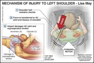
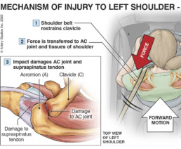
Shoulder Injury From Head-on Collision
Incident
This left-handed 56-year-old driver was involved in a head-on collision, causing soft tissue damage to her left shoulder as well as other injuries.
Purpose of illustration
This panel was created to show the mechanism of injury to her AC joint and supraspinatus tendon, resulting from a significant seatbelt force being suddenly applied to her shoulder during impact.
Rationale for litigation visual
The transmission of forces from the shoulder belt, to the clavicle and thereby to the bones and tissues of the shoulder, were key concepts to be communicated at trial. The illustration served to ‘translate’ the opinion of the biomedical engineer into a visual format, allowing for clear understanding.
Complications
Ms. May went on to experience persistent pain in her shoulder, reflecting the effects of inflammation and scarring of tendons, ligaments and bursa, with secondary mechanical effects.
Other visuals demonstrated
Additional illustrations showed more detailed findings of the shoulder pathology, as well as left knee injury concepts.
Outcome
Plaintiff’s lawyer, Norma Mayer, opined that “…the illustration was instrumental in resolving the file at mediation.”
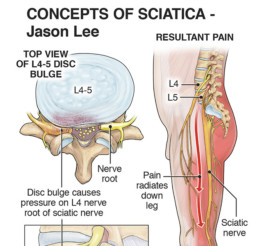
Trauma-Induced Sciatica
Illustrated Medicine: Case Study
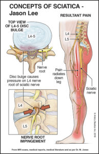
Trauma-induced sciatica
Incident
This 50 year-old seat-belted driver was rear-ended at highway speeds. He suffered a lumbar spine sprain/strain injury.
Purpose of illustration
This illustration exhibit was created to demonstrate the direct anatomical link between the trauma-induced L4-5 disc herniation diagnosed post-accident, and the resultant painful sciatica symptoms of shooting burning pain into his buttock and down his leg, making it difficult to stand and walk.
Rationale for litigation exhibit
Mr. Smith had previous surgery in this area and was susceptible to further injury. The jury needed to appreciate: details of the injured disc with herniation resulting from the impact forces; associated impingement of the L4 nerve root component of the sciatic nerve; and the resultant pain that radiated through his lower back and into his left lower limb in the L4 dermatome pattern.
Complications
The L4-5 disc underwent further degenerative changes. Mr. Smith had several injections for pain, and surgery was eventually required.
Other visuals demonstrated
Additional illustrations showed: details of the mechanism of injury; an overview of how disc herniation progresses to the point of nerve root impingement; and the associated decompression surgery that was performed.
Outcome
The case settled immediately before trial was to begin.
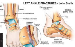
Trimalleolar Fracture-Dislocation
Illustrated Medicine: Case Study
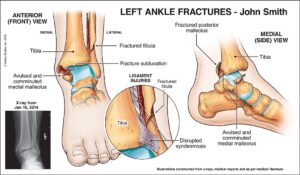
Trimalleolar Fracture-Dislocation
Incident
This 21-year-old construction worker slipped into a hole, sustaining a displaced/dislocated trimalleolar fracture that required open reduction internal fixation (ORIF). Osteoarthritis (OA) developed quickly in the joint due to the impact to, and disruption of, the articular cartilage.
Purpose of illustration
This panel was created to demonstrate the significant fracture-dislocation with associated injury to the syndesmosis (the distal tibia-fibula joint bound together by ligaments). The illustrations allowed litigation participants to fully appreciate the severity of the trauma and why OA ensued.
Rationale for litigation visual
Although x-rays demonstrated the fracture-dislocation, illustrations were required to translate the films into more readily understood images to show the full extent of trauma, including details of the syndesmosis injury.
Complications
Post-injury, this young man suffered pain on ambulation and at rest. His ankle joint demonstrated osteoarthritic degeneration within a year of the slip & fall. Some of the hardware and osteophytes required surgical removal. Further degeneration and associated surgeries were predicted for him.
Other visuals
Additional illustrations showed: the post-op appearance after ORIF surgery; hardware removal; future further osteoarthritic degeneration; and potential future ankle surgery – including debridement, fusion or replacement.
Outcome
This exhibit was created to allow the orthopedic expert to fully explain the extent of trauma this man sustained, and the significance of his intra-articular fractures. The illustrations were sent to defense and the case settled before trial.
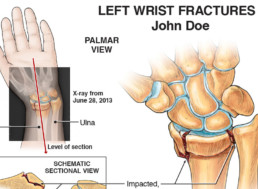
Intra-Articular Wrist Fracture
Illustrated Medicine: Case Study
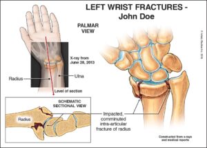
Intra-Articular Wrist Fracture
Incident
Mr. Jones was a 45-yo male driver involved in a head-on collision and required extrication.
Purpose of Illustration
This panel was created to show details of his left wrist injury, providing a visual translation of the radiology and medical reports. It also served to demonstrate the severity of the trauma-related issue(s), to address objections of pre-existing psoriatic arthritis and two prior MVA’s with unrelated sequelae.
Rationale for litigation visual
X-rays were too difficult for a jury to comprehend understand. Other visuals showed the surgery performed and degeneration concepts resulting from the significant damage he sustained to the articular cartilage in his wrist.
Complications
He was later shown to have early arthritic changes in his left wrist joints – based on x-ray findings and a bone scan. Experts opined that his wrist would likely continue to degenerate with osteoarthritis.
Other visuals demonstrated
Concepts of: concussion; lumbar spine injury/surgery; pain treatments; sciatica complications; and right foot issues including fractures, surgeries, and degeneration. A 3D-printed model showed details of his right foot fractures.
Outcome
The demonstrative evidence was created in time for mediation. Using the visuals, the lawyer walked through the medical issues in detail, linking them directly to the biomechanical engineering opinions. A significant settlement was reached shortly thereafter with the visuals showing the clear “facts” and informing the adjuster’s decision.
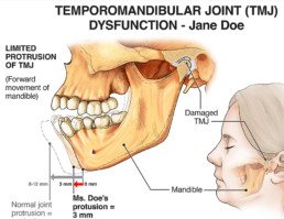
Complications from TMJ Trauma
Illustrated Medicine: Case Study
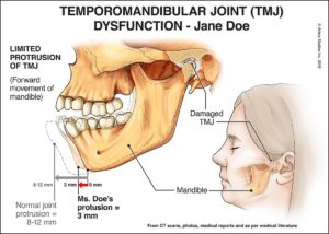
Complications from TMJ Trauma
Incident
This 15-year-old pedestrian was hit by a car on the left side. She flew onto the windshield, hit her head and landed on the pavement.
Purpose of illustration
This panel was intended to show findings noted by the dental expert associated with temporomandibular joint (TMJ) injury.
Rationale for litigation visual
TMJ pathology was the primary reason for the postural instability of her jaw and contributed to ongoing headaches.
Left-sided TMJ complications
She suffered pain with jaw opening and chewing, and had left-sided jaw pain at rest. Her jaw would lock closed, with instability of the joint.
Other visuals demonstrated
Additional illustrations showed issues of: mild TBI; R-sided hemianopia; sprain/strain injury to her neck and back; head/neck/back/shoulder pain; reduced range-of-motion of her C-spine; and vision problems.
Outcome
This visual was created to allow the dental expert to better explain an essential action of the mandible; it was to be used to educate litigation participants about key anatomical and functional issues of the jaw and TMJ; the case settled just prior to jury selection at the plaintiff’s pre-trial offer.